Bone Marrow Lab
At the bone marrow morphology lab, I usually view bone marrow slides or perform certain staining of slides (Maygrunwald Giemsa stain and iron stain). The staining in this lab is all done manually.
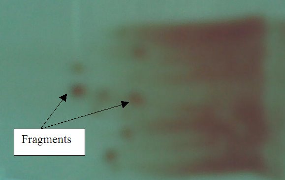
I also get to observe the bone marrow aspiration procedure. The doctors will perform the aspiration while the technologists collect the samples and place it into EDTA tubes or make on-site smears. There are usually 3 different smears made: Wedged smears, squashed smear and trephine imprint. The first 2 smears are routinely performed. These smears are somewhat different from the peripheral blood smear we have tried in school. Instead of using blood samples to smear, we are using fragments or marrow particles to smear.
For the wedged smear, the marrow sample is firstly placed on a clean slide. The slide is tilted to drain the blood, as only fragments are needed. Using a spreader (like we used in school), the fragments are ‘collected’. It is then smeared on a slide. For squashed smear, a few fragments are collected using the corner of the spreader. The spreader is then placed flatly on a glass slide, pushed up and smeared across. Fragments will be squashed in the center. Trephine imprint does not make use of marrow samples. Instead it uses the bone in the marrow (core plug). The bone piece is rolled in between 2 slides. An ‘imprint’ is formed. The trephine imprint is especially important if marrow samples cannot be collected due to dry tap (resulting in inability to do wedge and squashed smear). The slides will then be stained with Maygrunwald Giemsa stain and iron stain.

Wedged smear

Squashed smear

Trephine imprint
Internal QA in bone marrow morphology lab:
Bone marrow specimen handling
1)All specimens must be considered infectious. Thus technologists should wear lab coat and gloves. Specimens should be kept in a plastic specimen bags for transport.
2)spreaders used for smearing should be cleaned between cases. This is to reduce cross contamination.
Bone marrow sample processing
1) All smears and samples should be identifiable at all time.
2)A set of six well-made and labeled smears are selected for routine staining.
(a) choose 1 wedged smear with the most marrow particles for iron staining. A control slide with increased iron store should be stained at the same time.
(b) choose 1 peripheral blood film, 1 squashed smear, one trephine imprint and 2 wedged smears for Maygrunwald Giemsa stain.
3)Toxic wastes from special stains (cytochemical stain) should be disposed off carefully into separate designated containers for appropriate disposal.
4)All stained smears must be completely air-dried and mounted with cover slips using DPX. Air bubbles present should be removed.
Internal QC:
-Only positive controls are used in the lab.
-All control slides should be checked before the patient smears are examined.
-If control fails, identify the possible cause in the reagent preparation or staining procedure. Procedure should be repeated or a new lot of reagent should be prepared.
External QA in lab:
1) CAP survey on blood cell morphology from blood film and marrow.
2)RCPA survey on blood cell morphology from blood film and bone marrow.
3)CAP survey for cell morphology from CSF and body fluids.
4)CAP survey for blood parasites.
At the bone marrow morphology lab, I usually view bone marrow slides or perform certain staining of slides (Maygrunwald Giemsa stain and iron stain). The staining in this lab is all done manually.
I also get to observe the bone marrow aspiration procedure. The doctors will perform the aspiration while the technologists collect the samples and place it into EDTA tubes or make on-site smears. There are usually 3 different smears made: Wedged smears, squashed smear and trephine imprint. The first 2 smears are routinely performed. These smears are somewhat different from the peripheral blood smear we have tried in school. Instead of using blood samples to smear, we are using fragments or marrow particles to smear.
For the wedged smear, the marrow sample is firstly placed on a clean slide. The slide is tilted to drain the blood, as only fragments are needed. Using a spreader (like we used in school), the fragments are ‘collected’. It is then smeared on a slide. For squashed smear, a few fragments are collected using the corner of the spreader. The spreader is then placed flatly on a glass slide, pushed up and smeared across. Fragments will be squashed in the center. Trephine imprint does not make use of marrow samples. Instead it uses the bone in the marrow (core plug). The bone piece is rolled in between 2 slides. An ‘imprint’ is formed. The trephine imprint is especially important if marrow samples cannot be collected due to dry tap (resulting in inability to do wedge and squashed smear). The slides will then be stained with Maygrunwald Giemsa stain and iron stain.
Wedged smear

Squashed smear

Trephine imprint
Internal QA in bone marrow morphology lab:
Bone marrow specimen handling
1)All specimens must be considered infectious. Thus technologists should wear lab coat and gloves. Specimens should be kept in a plastic specimen bags for transport.
2)spreaders used for smearing should be cleaned between cases. This is to reduce cross contamination.
Bone marrow sample processing
1) All smears and samples should be identifiable at all time.
2)A set of six well-made and labeled smears are selected for routine staining.
(a) choose 1 wedged smear with the most marrow particles for iron staining. A control slide with increased iron store should be stained at the same time.
(b) choose 1 peripheral blood film, 1 squashed smear, one trephine imprint and 2 wedged smears for Maygrunwald Giemsa stain.
3)Toxic wastes from special stains (cytochemical stain) should be disposed off carefully into separate designated containers for appropriate disposal.
4)All stained smears must be completely air-dried and mounted with cover slips using DPX. Air bubbles present should be removed.
Internal QC:
-Only positive controls are used in the lab.
-All control slides should be checked before the patient smears are examined.
-If control fails, identify the possible cause in the reagent preparation or staining procedure. Procedure should be repeated or a new lot of reagent should be prepared.
External QA in lab:
1) CAP survey on blood cell morphology from blood film and marrow.
2)RCPA survey on blood cell morphology from blood film and bone marrow.
3)CAP survey for cell morphology from CSF and body fluids.
4)CAP survey for blood parasites.
posted by SyafiqaH at
10:30 PM
![]()

3 Comments:
Hi Syafiqah!
I didn't know that there was more than one way of performing a marrow smear. You mentioned that the Trephine imprint is performed when marrow samples cannot be collected due to dry tap. What do you mean by "dry tap"?
Also, I don't quite understand the part on external QA. In what way do the RCPA survey and CAP survey help in QA?
By Samantha, at 8:59 PM
Hello sam!
"dry tap" means that no marrow particles can be collected. Usually, the doctors will insert the aspiration needle into the bone and will "pull out" bone marrow fragments using a syringe. A "dry tap" is when nothing, or rather no marrow particles, comes out from the syringe. As theres no fragments, technologists will not be able to perform any smearing. That's where the imprint come in handy. Bone particle/ core plug will be used to "rub" between 2 microscopy slides. This way, cells from the bone surface will be somehow attached to the slide.
The RCPA and CAP will provide a sample or smeared slides. Technologists will perform the tests on these samples and will send the results to the CAP and RCPA, who will evaluate the results sent. The labs will then be informed of the outcome. It's like the external QA we learn in LMQA. If the results the lab sent in is close to that of other labs evaluated by the RCPA and CAP,this means that the lab is performing up to expectation and hence quality is assured.
If my explanation is still not very clear, do let me know. I'll try to clear your doubts.
=)
By SyafiqaH, at 8:21 PM
Hello Ms Chew
Generally, the control slides are used to determine if the staining done is acceptable. It is used to ensure that there is no false positive or false negative results of the sample slides.
For example, in the periodic acid schiff (PAS) stain, a normal blood film is used as control with every batch of stains. After staining, the control slides are firstly checked on the microscope. The neutrophils in the control slide must show an intense magenta/red cytoplasm stain. If it is not as stated, this means that the staining is not done properly. Hence, staining have to be repeated on a fresh set of slides.
By SyafiqaH, at 8:30 PM
Post a Comment
<< Home