Med Tech in Training

Sunday, July 23, 2006

External QA in Thalassemia Lab

The lab have 2 external QA:
1) College of American Pathologists (CAP)
2) RCPA (Australia-based)

The exercise is done 4 times a year for both CAP and RCPA. In the Thalassemia lab, CAP covers QA of the variant test, Hb-H test, Hb-A2 and Hb-F quantitation, sickling test and the alkaline and acid Hb electrophoresis test. The RCPA covers all tests except for the sickling test.
Unknown samples will be sent to the lab. The tests will be performed and results will be sent back to the the CAP and RCPA.

The CAP also covers disposal waste. Checks will be conducted to see if waste are disposed off accordingly. (e.g: Blood-stained equipments/objects disposed off in biohazard bins, used microscopy slides in sharp bins)

Hey all! Remember the entry I've written on HPLC (High performance liquid chromatography)? I have finally managed to understand the theory behind it, thanks to the notes the biotech students have compiled in one of their modules!

So I shall give you a rough idea of how this machine works and quality control procedures we perform. Just to refresh your memory, the purpose of HPLC is an analytical process utilizing special instruments designed to:
Separate, quantify and analyze components of a chemical mixture. This analysis that is being done is based on a chromatogram that is generated after the separation process.

So this is how it works

1. Samples which are stored in small bottles are introduced into this compartment that contains an automatic injector. This injector works by sucking up a certain quantity of fluid to be introduced into the system.

2. The sample is passed through the system in a high pressure solvent, aka mobile phase. This solvent will then carry the samples through a column packed with sorbents. This column is known as the stationary phase.

3. Okay, so here's the gist of how it works. The chemical mixture is separated based on their polarity. Depending on their polarity, the amount of interaction between the individual components and the mobile and stationary phase differ. The individual components that have the least amount of interaction with the stationary phase or the most amount of interaction with the mobile phase will exit the column faster.

For example, if the solvent we use is polar and the column is non-polar, polar components of the mixture will usually follow the solvent, and non-polar components will adhere to the column first, only leaving the column after the polar components have exited the column. Hence, there'll be a separation of individual components based on their polarity.

4. As the analytes exit the column, they can be detected by various means. The detector shines a light through the sample, and the light is detected and saved as an electrical signal. This siganl is sent to a computer which makes a graph of the data. This graph is a chromatogram which shows peaks of active compounds in the mixture. However, this chromatogram does not tell us anything about the identity of the compound. Hence, a known standard is often used to help in the identification.
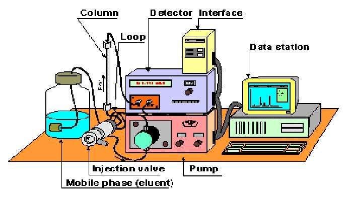


Basically, a standard refers to the active compound which you wish to identify. So after running the standard through the HPLC, a chromatogram will show up, showing only 1 major peak. By taking note of the retention time or the time it takes for the peak to show up, we'll be able to determine from the sample chromatogram which peak represents the studied active compound.
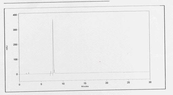

this is a chromatogram of the standard. It appears that the active compound shows up at a time frame of between 7 to 8 minutes.
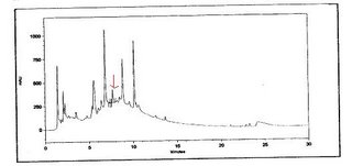

this is a chromatogram of the sample. the active compound is indicated by the arrow at a time frame of between 7 to 8 minutes.

Sunday, July 16, 2006

I’m assigned to be at the thalassemia lab for 3 weeks. The most common test requested is the Haemoglobin (Hb) electrophoresis test. This includes the alkaline cellulose acetate electrophoresis test, Hb-H screening test and the variant test.

The purpose of alkaline cellulose acetate electrophoresis is to screen for Hb variants (Hb E, Hb S, Hb F, Hb A). The variation in the amino acid content of the different Hb causes variation of net charge of each Hb type. This hence determines their rate of mobility. When run, the Hb bands will appear accordingly. Any variants detected is followed up with acid gel electrophoresis (to distinguish Hb C) or other tests.

QC of alkaline cellulose acetate electrophoresis:
1) Known Hb A,F,S and E controls are run on the first well of each gel plate.
Hence, this acts as a reference to determine what variant the bands represent.


Applicator used to "pick up" the samples.


The wells where the samples will be pipetted into. the applicator will then be placed into the wells to "pick up" samples


Electrophoresis buffer tank. The bigger columns on the top and bottom is filled with fixed amount of supre-heme buffer. The gel strips will then be placed on top of the centre columns.


An example of an alkaline cellulose acetate electrophoresis strip. The bands are as indicated by the arrows (controls).

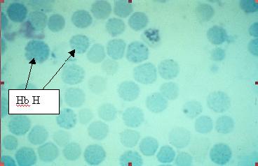
The Hb-H test is performed to screen for presence of Hb-H inclusion bodies.In this test, 2 drops of brilliant cresyl blue stain is added to 100 microlitres of blood. After incubation period of 1 hr at 37 degree celcius, samples are left to cool before smears are made for microscopy viewing. Presence of Hb-H is indicated by “golf-like” cells.

smearing of slides to screen for Hb-H. Notice that the colour of the smear is green due to the stain. The stain is bluish in colour at first. But it turns green when dried.


Picture taken from http://www.cuhk.hk/med/paf/slides/hematolo/xv-18.htm

The variant test (automated) is used to determine the percentage of Hb A2 and F. The machine used is the Bio-Rad variant beta-thalassemia short programme.

Monday, July 03, 2006

Week One

Hey guys! Farhana here. I’m currently attached to the Virology Lab. The Virology lab is divided into 3 main sections, namely the Serology, Isolation and Immunofluorescence(IF) section. Under the Isolation Section, there would be the Tissue Culture Lab and Virus Isolation Lab. At the moment, Li Jun and I are attached to the IF section. As the name implies, in the virology lab, we would be dealing with (duh!)….VIRUSES!!! Oooo… Sounds dangerous rite? Haha… Anyways, in the Virology Lab, we would be involved in the isolation, growth and testing for viruses such as Herpes Simplex Virus(HSV) and etc.

On the first day of SIP, we were given a briefing on the rules and safety and also given a mini tour of the Labs and introduced to the staff there. The first two days, we didn’t really get to do much except read the manuals and observed some of the procedures in the Tissue Culture Lab because the IF med techs were busy. In the Tissue Culture Lab, they basically do the same things that we did in MCT practicals. We all already know that viruses need a living host to survive, right? Yes? *Nodding* So clever! So, anyway, the purpose of the Tissue Culture Lab is to grow and maintain the cell lines to support growth of the viruses. Yes, that would mean that the use of trypsin, and MEM media and cell lines such as Hela would be involved. Sounds familiar, right?

Anyway, the first week in the IF Lab has been quite fun actually. Haha… because the med techs there had aliquoted for us some extra patient specimens and let us try out some of the tests. The first test that we got to try is the Nasopharyngeal Aspirate(NPA) test. In this test, we first take a NPA specimen(Btw, the NPA specimen can be quite disgusting. Some are watery, some bloody, some greenish, some spilled, some are plain.... *puke*) and add saline buffer(PBSA), vortex and then centrifuge to wash it. The washing step is done twice to ensure that the mucus in NPA would break up and release the cells trapped in them. If the sample is bloody, cold distilled water is added to lyse the RBC and PBSA is then added to stop the lysing reaction. When the samples are ready, they can then be spotted onto the wells on the slides(like in picture below) and then fixed in acetone.


After fixation, we would then add monoclonal antibodies(eg anti-influenza Monoclonal Ab), incubate and wash and then add the conjugate, incubate and then wash and left to air dry. After drying, we can then mount the slides. And TADA!! We can then view the slides. Since we’re in an Immunofluorescence Lab and are doing an Immunofluorescence Assay, that would mean that we get to use the -----> Immunofluorescence microscope. Haha! I think the great thing about being attached to the IF lab is getting to use the IF microscope cause I think that the cells look really cute under the microscope. Haha… The positive cells are stained apple-green while the negative ones are stained a dull-red colour. Actually, it’s quite hard to know whether the slides are really positive for the viruses cause debris and dirt may take up the dye and show up as a yellowish green fluorescence. Anyway, below is a picture of how a slide looks under the IF microscope.


Oh and while doing the slides for the patient's specimens, we also need to prepare and test a control slide containing the positive and negative control. This would help confirm the validity of the results of the patient's samples by ensuring that there isn't any cross-contamination between the different samples and the reagents.

I guess I’ll stop here for now. Will update ya’ll soon… Bubbye!

Sunday, July 02, 2006

Lab safety - Fire Safety

On the first day of attachment, we were told to read up on certain reading materials. For this entry, I will recall the main important points stated in the company’s basic fire safety handbook.

-->The company make use of “RACES” in cases of fire. R- rescue, A-alarm, C-call, E- extinguisher, S-shut (shut down gas supply and close doors to contain the fire and smoke).


-->There are 3 types of extinguisher:
1)Water extinguisher (red colour): suitable for ordinary fires
2) Dry chemical extinguisher (blue colour): for all types of fire
3)Carbon dioxide extinguisher (black colour): for liquid or electrical fires


-->How to operate fire extinguisher:
1)Pull safety device
2) Aim nozzle
3) Press trigger/lever


-->Basic fire safety precautions:
1) No smoking in hospital’s compound.
2) Keep flammable liquids in proper containers and store them separately from other combustible materials.
3) Access to fire fighting equipment such as the extinguisher should not be blocked.
4) Electrical and gas appliances should be turned off when not in use or after use.
5) Potential fire hazards should be promptly reported to safety personnel.


-->Procedures in case of fire:
1) Alert other staff of the fire
2) Break the nearest fire alarm call point
3) Inform Fault Reporting Centre of the fire
4) Try to put off the fire by using the fire extinguisher
5) Evacuate the place

Besides reading about fire safety, we were also shown where the fire alarms and extinguishers are located. We were also shown the exit route we should take in case there is fire.

During the week, I was assigned to be at the sample reception, whereby blood and urine samples are collected via the telelift, pneumatic tube or by hand. The samples were then sorted according to types of tests to be carried out. Next it is labelled and sent to the routine laboratory.

In maintaining or ensuring desired quality of the results, we must:
1) Check if the amount of blood collected is sufficient. If there is inadequate amount of blood, the amount of EDTA may be higher compared to the blood itself. This will lead false results.
2)Check if the name of the patient stated in the request form coincide with that of the sample tube. This is to ensure that the results printed will be for the correct patient.
3)Check if the blood has clotted or lysed to prevent false results.
Samples should be rejected if it fails to “pass” one or more of the above 3 points.

Saturday, July 01, 2006

Hi all! So how has your week been at your attachment sites?

As some of you may know, I'm attached to a research lab in school. Currently, I'm researching on traditonal chinese medicine for both my major project and the research competition organised by NUS.

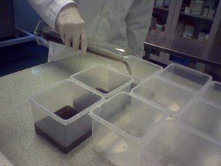
As for my SIP, I have just started it on friday and my scope is on plant tissue culture. In plant tissue culture, plants are actually grown in agar medium. (can you believe that?! I was quite amazed when i first saw it) So what I basically did on friday was subculturing of plants that have overgrown into new agar media. This was done by cutting away the leaves and leaving the trimmed part of the stem and roots behind to be immersed into the agar.


The container on the right shows the subcultured plant tissue into the agar. The agar contains activated charcoal that helps to absorb waste, which explains why the agar is black


Okay, enough of what I've been doing. From what I have observed so far, the lab is managed by a rather tight security system. From the entry into the main lab to the other specialized labs (other rooms within a lab such as the chemistry lab, plant tissue culture lab, TCM research lab), a security card is needed to gain access to these various labs. It works by tapping the card against this scanner that would verify if access is allowed into a particular lab. Depending on the card that is being used, it will only allow access to certain labs. This is practiced to minimize unnecessary movement by students into labs which are not intended for their use.

Another feature of quality control is traceability. This is practiced through recording the time and date of use of equipment (such as centrifuges) as well as the name of the person using the equipment into a log book. This is done so that if any fault arises in the machine, we'll be able to trace back and find out the identity of the person who was accountible for the error.

Also, to ensure the quality of reagents that are used in the lab, once a new reagent bottle is opened, the date of use should be recorded on the bottle or pack so that we'll be able to determine the freshness of the reagent.

Since I'm using herbs for my MP and the research competition, the consistency of these herbs have to be ensured by performing HPLC or high performance liquid chromatography. I'm still unsure of how the whole HPLC machine works, but all I know is that the machine operates by separating the components of the herb and generating a chromatogram with peaks. These peaks indicate active compounds in the herb and by analyzing the profile of the chromatogram between 2 or more batches of herbs, we'll be able to know the consistency of quality between these batches. So if the profile appears to be similar, the herbs can be used. However, if the profiles appear different, then further analysis has to be done to find out why the quality of herbs differ. This could be due to the inconsistent extraction process or some other reasons.