Med Tech in Training

Wednesday, November 01, 2006

Finger / Heel Prick

I have observed both the finger prick and heel prick procedure. Heel pricks are usually done on premature babies. The blood taken from both the finger and heel pricks are used to perform the full blood count (FBC) test. In addition for premature babies, the reticulocyte count test may also be requested.

The lancet or needle used for the pricking is different to those we used during one of the haemotology practical session in school. The lancet used in this company has a round base and can be positioned in any way. In contrast the lancet we used in school has a rectangular base and the lancet must not be positioned parallel to the fingerprints.

There are several points that should be noted when performing the pricking procedure:
1) Gloves should be put on before pricking procedure
2) the gloves should be changed after pricking each patient. This is to reduce contamination.
3) Wipe finger or heel with alcohol swab first and allow the alcohol to evaporate before pricking.
4) Wipe the first drop of blood with a gauze before collecting subsequent drops.
5) When 'squeezing or milking' for blood, press the fingers and let go, before pressing again.

Thursday, October 19, 2006

Automation and its advantages

I have learnt quite alot about automation through this attachment process. By experiencing automation first hand, i have a deeper understanding on automation and its uses, which we have learnt in chapter 1 of LMQA.

In the lab, several tests are performed using an automated method. This includes Full Blood Count (FBC) test and coagulation test. The samples for FBC are run using either the Advia, Sysmex or LH FBC analyser while the coagulation samples are run using the Sysmex CA-1500 machine. One clear advantage is that by using automated means, the samples are run at a faster rate. For example, if PT/APTT test for coagulation is performed manually, alot of time is required as one staff can only handle one sample at a time. In contrast, several samples can be placed in the machine and run at any one time, although the test will still be carried out one by one. Furthermore, manual method may bring about inconsistency as a person may only notice the clot a few seconds after it happens.

In the routine haematology lab i'm attached to, staining of slides are also performed automatically using the HemaTek stainer. If staining is done manually, it will take 6 mins for the staining process alone and a further 3-5 mins for the slides to dry. On the other hand the approximate rate of staining using the HemaTek stainer is 1 slide/ min. One will only need to place the slide on a conveyor-like space and the slides will be automtically stained, rinsed and dried.


HemaTek Stainer


Some people may think that human resource is needed much less with the use of automated machine. However, this is not necessarily true. Machine only does test and produces result for us. It is not able to analyse and troubleshoot. For example, the Advia FBC analyser will only alert technologists by producing (*)s on the result worksheet. The technologist's job is to identify the fault and correct it. For example when starting up the analyser, if the control did not fall within the given range, the technologist have to figure out what is wrong (e.g: check if the reagent is expired).

Steps to follow when the result worksheet is printed:
1) Make sure that the MCHC is less than 37 and more than 32, HCT is about 3 times more than Hb, difference between MCHC and CHCM is within 2. If not, repeat test.
2) check for panic range :
- Platelet less than 20 or more than 800 x10(9)/L
- WBC less than 1 or more than 50 x10(9)/L
- Hb is less than 5 or more than 22 g /dL for females or 20 g/dL for males.
If there are any panic ranges, call the wards to inform the doctor of the patient's platelet/wbc/Hb levels.
3)If the platelet level is below 140 x10(9)/L, check blood sample for presence of blood clot. If there is a blood clot, reject the sample and request for a repeat.

Sunday, October 01, 2006

Overview of tests done in urinalysis lab

(a) Dipstick – 2 types
1) five-patch test strip to determine quantity of pH, protein, glucose, ketone bodies and blood in urine.
2) 10-patch test strip for glucose, bilirubin, ketone, specific gravity, blood, pH, protein, urobilinogen, nitrite and leukocytes in urine.

The dipstick test is performed routinely. However, it is only done if the sample is that of an outpatient or patient with renal disease. Usually, only the 1st dipstick consisting of 5 tests is used. The 10-patch test stip is used only if the specific tests, such as bilirubin and urobilinogen, are requested.

(b) Microscopic analysis (using KOVA glasstic 10 with grid slide)
This is used to determine or identify cells that may be found in urine samples.9 microlitre of urine is pipetted into a notch on the KOVA glasstic slide chamber. The slide consist of grid lines that is used to count the number of specific cells. Examples of possible cells found in urine includes:
1) White blood cell
2) Red Blood Cell
3) Casts
4) Crystals such as cystine and triple phosphates
5) Microorganism (bacteria, yeast, spermatozoa)

An example of a KOVA slide. (taken from http://www.mlo-online.com/articles/0104/mlo0104prodfocus.htm)

- Internal QA -
1) The tests should be performed within 2 hrs of collection. The chemical composition of urine (e.g: pH,glucose and bilirubin) may change after 2 hrs. Furthermore, any formed elements such as crystals that are actually present in the urine, may begin to deteriorate and become unrecognizable.

2) For routine urinalysis, the preferred type of urine specimen is that of a midstream clean catch. This is so as that type of specimen reflects the urine composition in the bladder and the kidney

3) Dipstick Testing
(a) The dipsticks must be protected from moisture, light, heat and volatile chemicals as they deteriorate easily.
(b) They also deteriorate with time and hence should not be used after the expiration date.
(c) Each dipstick bottle is tested routinely (daily, before dipsticks are used) for results against known commercial controls. Each of the chemical parameters of the dipstick is evaluated and the results recorded (e.g: protein 2+, pH 6.5) By doing this, the technologist performance can be compared on a daily basis and the reactivity of the strips can be checked against known standards.
(d) Only a few strips should be removed at a time and the container should be closed tightly immediately after taking out the strips.
(e) The amount of time required for the colour reaction to develop on the strip varies with each test (e.g: 2 minutes for leucocytes). A timing device (stopwatch) is used and the manufacturer’s timing instructions should be followed strictly. The strip should be held close to the colour chart in adequate lighting when reading the results.

Sunday, September 10, 2006

WEEK 5 & 6

It’s been a month already, Li Jun and I have now moved on to the Isolation Section of the Virology Lab. The Isolation section consists of the Tissue Culture Lab and the Viral Isolation Lab. The Tissue Culture(T/C) Lab is where the med techs grow and prepare cell lines such as Hela and MDCK that act as hosts for viruses in shell vials, flasks and tubes. The Isolation Lab is the site where the med techs would inoculate samples of patients with suspected viral infections into the cell lines and culture and observe the cultures for over a period of time. Cultures grown in the tubes would be viewed for cytopathic effect(CPE) and tests such as the neutralization test or acid tests would be done to confirm the results. Culture tubes for some viruses such as HSV are sent to the Immunofluorescence Section for Ag IF detection.

Quality Control
In the Tissue Culture Lab, when we prepare the media and reagents, etc. We need to record down the batch details in the Quality Control Record for Media and Reagents. Details recorded include the date of preparation & expiry, result of sterility check, date of use, initial of the med tech responsible for the preparation of the reagent or media.

Sterility test
For every batch of media or reagent prepared, a sterility test must be done.
1) Inoculate 3ml of prepared medium/solution to 1 tube of Nutrient and 1 tube of Sabouraud’s broth.
2) Incubate Sabouraud’s tube at Room Temperature and Nutrient Broth tube at 37C for 7 days, observe daily.
- Discard any aliquot corresponding to tube which shows signs of growth.
- The sterility test is done to ensure that the reagents prepared are not contaminated with bacteria(Nutrient broth) or fungi(Sabourad’s broth) so that the cell lines would not be contaminated.

During our attachment in the Isolation Lab, Li Jun and I were given a sample each to isolate and observe. I was provided with a throat swab sample from a patient who was suspected of having Hand, Foot and Mouth Disease(HFMD). The test required for this particular sample is the Enterovirus culture. Before we start inoculating the specimens, we first need to process the sample by allocating a lab number for the sample and recording down the time and date the specimen was received and also include the initials of the person who was responsible for the receipt of the specimen. We then selected the tubes/shell vials of cell lines needed for inoculation of the sample. For the throat swab sample, a pre-adsorption is done. Pre-adsorption is believed to enhance entry of viruses into the cells. The medium is first removed from the cell line tubes and the 0.3 ml of the specimen is inoculated in to the tube and the tubes incubated at 36C for 1 hour after which, the tubes would be re-fed with medium and incubated and observed daily over 21 days and the results recorded in a specimen worksheet.

The tubes are read daily so as to detect any Cytopathic Effect(CPE) that may be present. When viruses multiply in the cell lines, they usually produce changes in the growing cells. This change is called the CPE of the virus and may be characteristic of the particular virus. A preliminary identification can be made by the appearance of the CPE. However, we must always confirm the result by performing a more specific test such as an immunofluorescence assay.

WEEK 7 & 8
During the period of observation, the tubes need to be maintained by regularly changing the media when it has become too acidic(yellow) and also the tubes(except for diploid cell lines) need to be repassaged every 7 days.

Repassaging of culture tubes
1) Scratch the cells off the inside wall of the tubes using a bent pipette.
2) Vortex the tubes to break up clumps of cells.
3) View the tubes under the microscope to ensure that most of the cells have detached.
4) Incoulate 0.2ml of the cells into a new cell line tube.

Repassaging is done to prevent overgrowth and overcrowding of the cells.

During the last week of observation, I got a shock when I saw there was fungi growing in the media and the inner walls of 2 of my culture tubes. I immediately informed the med tech there and she taught me how to filter the contaminated tubes.

Filtering of contaminated tubes
1) Freeze and thaw the tubes 3 times.
2) Filter the culture using a 0.2 Um millipore filter.
3) Inoculate 0.2ml of the filtered cells into a new cell line tube.

Eventhough my tubes got contaminated, I wasn’t upset about it because I realized that I actually benefited from it. Before the incident, I had observed a med tech there perform the filtering of contaminated tubes. However, she was too busy to explain the steps in detail. This incident has allowed me to perform the filtering myself and thus, I get to learn and remember the steps better and I also understand the rationale behind each step.

Sunday, August 27, 2006

Bone Marrow Lab

At the bone marrow morphology lab, I usually view bone marrow slides or perform certain staining of slides (Maygrunwald Giemsa stain and iron stain). The staining in this lab is all done manually.

I also get to observe the bone marrow aspiration procedure. The doctors will perform the aspiration while the technologists collect the samples and place it into EDTA tubes or make on-site smears. There are usually 3 different smears made: Wedged smears, squashed smear and trephine imprint. The first 2 smears are routinely performed. These smears are somewhat different from the peripheral blood smear we have tried in school. Instead of using blood samples to smear, we are using fragments or marrow particles to smear.

For the wedged smear, the marrow sample is firstly placed on a clean slide. The slide is tilted to drain the blood, as only fragments are needed. Using a spreader (like we used in school), the fragments are ‘collected’. It is then smeared on a slide. For squashed smear, a few fragments are collected using the corner of the spreader. The spreader is then placed flatly on a glass slide, pushed up and smeared across. Fragments will be squashed in the center. Trephine imprint does not make use of marrow samples. Instead it uses the bone in the marrow (core plug). The bone piece is rolled in between 2 slides. An ‘imprint’ is formed. The trephine imprint is especially important if marrow samples cannot be collected due to dry tap (resulting in inability to do wedge and squashed smear). The slides will then be stained with Maygrunwald Giemsa stain and iron stain.

Wedged smear


Squashed smear


Trephine imprint

Internal QA in bone marrow morphology lab:
Bone marrow specimen handling
1)All specimens must be considered infectious. Thus technologists should wear lab coat and gloves. Specimens should be kept in a plastic specimen bags for transport.
2)spreaders used for smearing should be cleaned between cases. This is to reduce cross contamination.
Bone marrow sample processing
1) All smears and samples should be identifiable at all time.
2)A set of six well-made and labeled smears are selected for routine staining.
(a) choose 1 wedged smear with the most marrow particles for iron staining. A control slide with increased iron store should be stained at the same time.
(b) choose 1 peripheral blood film, 1 squashed smear, one trephine imprint and 2 wedged smears for Maygrunwald Giemsa stain.
3)Toxic wastes from special stains (cytochemical stain) should be disposed off carefully into separate designated containers for appropriate disposal.
4)All stained smears must be completely air-dried and mounted with cover slips using DPX. Air bubbles present should be removed.

Internal QC:
-Only positive controls are used in the lab.
-All control slides should be checked before the patient smears are examined.
-If control fails, identify the possible cause in the reagent preparation or staining procedure. Procedure should be repeated or a new lot of reagent should be prepared.

External QA in lab:
1) CAP survey on blood cell morphology from blood film and marrow.
2)RCPA survey on blood cell morphology from blood film and bone marrow.
3)CAP survey for cell morphology from CSF and body fluids.
4)CAP survey for blood parasites.

Monday, August 21, 2006

Hmmm... These posts are long overdue...

Week 2

In the Immunofluorescence Lab, we were given daily tasks of recording the temperature of the lab and instruments such as the refrigerator and incubator and also the calibration of the counter analyzer. The recording of the temperature these instruments is necessary so as to ensure that the reagents used for the diagnostic tests are stored at the right temperature while the calibration of the counter analyzer is done to ensure that the results produced are accurate and consistent.

Calibration of the counter is done every morning before actual patients’ samples are analyzed. Calibration is done by running 3 blood controls of different values: Abnormal Low, Normal and Abnormal High. The control values yielded by the counter should fall within the ranges provided. If the values are out of range, the controls should be run again. To prevent false high or low results of the controls, after removing them from storage in the fridge, the controls should be left to warm up to room temperature and should be mixed well first. However, the blood control tubes should not be shaken too vigorously as it would cause bubbles. The controls should also be capped back tightly to prevent clotting of the blood and stored back in the fridge immediately after use. After the controls are run, the results would then be printed out and kept in the QC file. The counter would then be ready for use.

On Tuesday, while calibrating the counter analyzer, an unfortunate incident happened. One of the control tubes broke, spilling its content onto the floor. The medical technologists there cleaned up the blood spill according to the protocol set for Biological Spills.

Biological Spill
1) Warn co-workers.
2) Contain or cover the spill with paper towels.
3) Cover spill with 10% bleach or 1% Virkon, pouring slowly from the outside in.
4) Let the spill soak for 10 mins.
5) Pick up the soaked paper towels and dispose then in the yellow biohazard bag.
6) Inform Lab Safety officer.

Eventhough the incident was an unfortunate one & had caused a disruption in the daily workflow of the lab, I think that both, us the students and the medical technologists there learnt something from it. For us students, we got to observe an actual biological spill and the steps done to clean it up. For the med techs there, they now know that they should aliquot some of the control into another tube and keep them as a back-up so that if a similar incident were to occur, they would have another set of controls and they can continue calibrating the counter and continue with the diagnostic tests.

Week 3

Besides performing the daily recording of the temperature of the lab and instruments and calibration of the counter analyzer, we learnt and performed the staining of mycoplasma and detection of Measles Antibody.

Cell lines grown in the lab are tested for mycoplasma contamination. Cell lines with passage numbers #0, 1, 2, 7 are cultured in 1 ml of medium void of any antibiotics in shell vials containing cover slips.

Bisbenzimide staining of mycoplasma
1) Make a 1/100 dilution from stock( 50 ug/ml) with absolute methanol.
2) Remove growth medium from the coverslip and wash with PBSA twice.
3) Rinse the cells once with working Bisbenzimide(0.5 ug/ml), then cover cells with about 0.5 ml of working Bisbenzimide.
4) Incubate the coverslip at 36˚C for at least 15 mins.
5) Pour off the bisbenzimide solution & rinse the cells with absolute methanol.
6) Mount and read.

Like any other tests done in the lab, positive and negative controls should also be included in each run.

In school, during Mammalian Cell Tech lectures and practicals, we learnt about how we should always practice aseptic techniques and also view our cell lines cultures daily so that if there are abnormalities in cell morphology or medium due to contamination, we could notice it immediately. We also learnt that unlike bacterial contamination, mycoplasma contamination does not cause turbidity in the medium and is thus harder to detect. Although we learnt these stuff in school, we didn’t have the opportunity to carry out the actual detection of possible mycoplasma contamination in the cell line cultures. So, I think it’s great that we get to do something related to what we have learnt.

Besides mycoplasma staining, we also performed the detection of Measles IgM and IgG.

Anti-Measles IgM IF
1) Treat serum with Gullsorb to give a final dilution of 1/10 as follows:
5 uL serum: 1 drop Gullsorb
2) Add the patient sera and positive and negative control sera to the wells on the Measles antigen slides.
3) Incubate the slides at 37˚C for 90 minutes in a moist chamber.
4) Rinse the slides twice with a stream of PBSA and immerse slides in 200 ml of PBSA and shake at 50 rpm on a shaker for 20 minutes.
5) Shake off excess PBSA from slides and add the Anti-Human IgM FITC conjugate to all wells.
6) Incubate the slides at 37˚C for 30 minutes in a moist chamber.
7) Rinse the slides twice with a stream of PBSA and immerse slides in 200 ml of PBSA and shake at 50 rpm on a shaker for 20 minutes.
8) Allow slides to air dry.
9) Mount and read slides.

The protocol for the Measles IgM and IgG tests are similar except that for the IgM test, we need to add gullsorb instead of the PBSA added in the IgG test. Gullsorb is added to dissolve the IgG found in the sera. This is important as IgG can interfere with the assay for IgM in 2 ways:
1) by competing with specific IgM for substrate binding sites
2) by forming immune complexes with rheumatoid factor which can mimic IgM, becoming a reactive site for the conjugate, thus producing an IgM false positive result.

For the first practice for the Measles Ab test, everything went smoothly and we were successful in getting the correct results. However, for the 2nd practice, we accidentally added Antigen conjugate instead of the Antibody conjugate meant for the Ab test. Haha… ya, we were very blurrrr and silly…But luckily, we realized our mistake and washed the slides thoroughly and proceeded to add the correct reagent. Despite our mistake, we still managed to get the correct results. We learnt that it is because the Ab conjugate consists of an anti-human Ab whereas the Ag conjugate contains anti-mouse Ab. Thus, the Ab did not react and interfere with assay.

Week 4

Quality Assurance

External quality assessment programmes in which the laboratory participates are listed in the virology quality manual.

They consist of the following:
College of American Pathologist
i. Viral antigen survey
ii. Diagnostic immunology survey for rubella antibody
iii. Viral culture survey
iv. Viral markers genes 1 and 2 survey
v. Viral antibody survey
vi. Hepatitis C viral load survey
vii. Nucleic acid amplification survey
viii. C. trachomatis and N.gonorrhoeae (NAA) survey

WHO Immunology laboratory
National hepatitis B Quality Assurance Programme for hepatitis B surface antigen and antibody

WHO poliomyelitis Eradication Proficiency Testing for enterovirus isolation

Royal College of Pathologist of Australia Microbiology QAP
i. Antenatal serology
ii. Vaccine preventable disease serology
iii. Viral serology
iv. Trial module

National Serology reference laboratory, Australia

Centre of disease control and prevention (HIV-1) Antibody testing

Sunday, July 23, 2006

External QA in Thalassemia Lab

The lab have 2 external QA:
1) College of American Pathologists (CAP)
2) RCPA (Australia-based)

The exercise is done 4 times a year for both CAP and RCPA. In the Thalassemia lab, CAP covers QA of the variant test, Hb-H test, Hb-A2 and Hb-F quantitation, sickling test and the alkaline and acid Hb electrophoresis test. The RCPA covers all tests except for the sickling test.
Unknown samples will be sent to the lab. The tests will be performed and results will be sent back to the the CAP and RCPA.

The CAP also covers disposal waste. Checks will be conducted to see if waste are disposed off accordingly. (e.g: Blood-stained equipments/objects disposed off in biohazard bins, used microscopy slides in sharp bins)

Hey all! Remember the entry I've written on HPLC (High performance liquid chromatography)? I have finally managed to understand the theory behind it, thanks to the notes the biotech students have compiled in one of their modules!

So I shall give you a rough idea of how this machine works and quality control procedures we perform. Just to refresh your memory, the purpose of HPLC is an analytical process utilizing special instruments designed to:
Separate, quantify and analyze components of a chemical mixture. This analysis that is being done is based on a chromatogram that is generated after the separation process.

So this is how it works

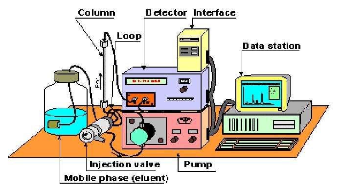
1. Samples which are stored in small bottles are introduced into this compartment that contains an automatic injector. This injector works by sucking up a certain quantity of fluid to be introduced into the system.

2. The sample is passed through the system in a high pressure solvent, aka mobile phase. This solvent will then carry the samples through a column packed with sorbents. This column is known as the stationary phase.

3. Okay, so here's the gist of how it works. The chemical mixture is separated based on their polarity. Depending on their polarity, the amount of interaction between the individual components and the mobile and stationary phase differ. The individual components that have the least amount of interaction with the stationary phase or the most amount of interaction with the mobile phase will exit the column faster.

For example, if the solvent we use is polar and the column is non-polar, polar components of the mixture will usually follow the solvent, and non-polar components will adhere to the column first, only leaving the column after the polar components have exited the column. Hence, there'll be a separation of individual components based on their polarity.

4. As the analytes exit the column, they can be detected by various means. The detector shines a light through the sample, and the light is detected and saved as an electrical signal. This siganl is sent to a computer which makes a graph of the data. This graph is a chromatogram which shows peaks of active compounds in the mixture. However, this chromatogram does not tell us anything about the identity of the compound. Hence, a known standard is often used to help in the identification.


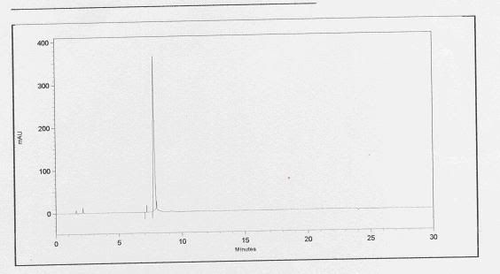
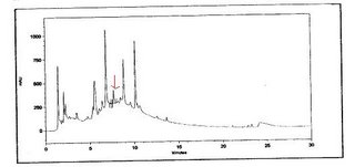
Basically, a standard refers to the active compound which you wish to identify. So after running the standard through the HPLC, a chromatogram will show up, showing only 1 major peak. By taking note of the retention time or the time it takes for the peak to show up, we'll be able to determine from the sample chromatogram which peak represents the studied active compound.


this is a chromatogram of the standard. It appears that the active compound shows up at a time frame of between 7 to 8 minutes.


this is a chromatogram of the sample. the active compound is indicated by the arrow at a time frame of between 7 to 8 minutes.